Anatomy Of Chest Area : 1 Anatomy Of The Arm And Chest Used With Permission Of N Moureau Download Scientific Diagram - General anatomy neuroanatomy head and neck anatomy thoracic anatomy abdominal and pelvic anatomy spinal anat.
Anatomy Of Chest Area : 1 Anatomy Of The Arm And Chest Used With Permission Of N Moureau Download Scientific Diagram - General anatomy neuroanatomy head and neck anatomy thoracic anatomy abdominal and pelvic anatomy spinal anat.. The sternum, or breastbone, is a flat bone at the front center of the chest. The electrical impulse then travels to an area of cells at the bottom of the right atrium, between the atria and ventricles, called the atrioventricular, or av, node. Browse 2,552 female chest anatomy stock photos and images available, or start a new search to explore more stock photos and images. An opacity (a normally dark area appears light on the image) a density (a. These areas are also known as the hidden areas.
The electrical impulse then travels to an area of cells at the bottom of the right atrium, between the atria and ventricles, called the atrioventricular, or av, node. Identify the basic anatomy seen on a chest radiograph. Shortness of breath comes on with physical activity sometimes with light activity or even if talk too much. An opacity (a normally dark area appears light on the image) a density (a. In particular, the right side of the chest is home to several structures including the right side of the heart, the three lobes of the right lung, the ascending aorta, the pulmonary blood vessels,.
Chest Anatomy Order from presentationgroup.com Anatomy of chest area : System respiratory respiratory organs of human body digestive and respiratory system medical chest internal structure of human body medicine body lungs biology intestines stomach anatomy torso human internal. Most chest bone fractures are due to trauma from a car accident. Find the perfect chest anatomy stock photo. The best upper chest workout will. Experts would obtain a preliminary supine scout radiograph of the chest with lead markers at 2cm intervals to localize the area of interest. It is therefore important to look at every part of the image in a careful and systematic way. Download my two educational text books for free using this link:
Identify the basic anatomy seen on a chest radiograph.
In the human body, the region of the thorax between the neck and diaphragm in the front of the body is called the chest. General anatomy neuroanatomy head and neck anatomy thoracic anatomy abdominal and pelvic anatomy spinal anat. The corresponding area in an animal can also be referred to as the chest. The bottom of the manubrium forms a joint with the uppermost portion of the body of the sternum known as the manubriosternal joint. The shape of the chest does not correspond to that part of the thoracic skeleton that encloses the heart and lungs. Fractures in the manubrial area are the second most common. The diaphragm forms the upper surface of the abdomen. The internal layer is noncontinuous around the inner surface of the chest wall and comprises the innermost intercostals, the subcostals, and the. The chest anatomy includes the pectoralis major, pectoralis minor & serratus anterior. He has been also having pain in the throat area and feeling very fatigue and weak. Chest a man's chest — like the rest of his body — is covered with skin that has two layers. Anatomy of the upper chest area. Thorax surface anatomy 4 edition / chest a man's chest — like the rest of his body — is covered with skin that has two layers.
Anatomy of chest area : Download my two educational text books for free using this link: Anatomy of the chest and shoulder, anatomy of the chest organs, anatomy of the chest wall, anatomy of the chest wall and pleura, anatomy of upper chest area, human. Most chest bone fractures are due to trauma from a car accident. The frontal chest radiograph and axial chest ct images are viewed as if looking at the patient, with the patient's right side on the viewer's left.
Thorax Wikipedia from upload.wikimedia.org The best upper chest workout will. Using seat belts prevent most of these types of injuries. The pectoral region is located on the anterior chest wall. It is therefore important to look at every part of the image in a careful and systematic way. System respiratory respiratory organs of human body digestive and respiratory system medical chest internal structure of human body medicine body lungs biology intestines stomach anatomy torso human internal. You can observe for it and. Huge collection, amazing choice, 100+ million high quality, affordable rf and rm images. The corresponding area in an animal can also be referred to as the chest.
Gurmukh singh answered 49 years experience pathology sternal angle:
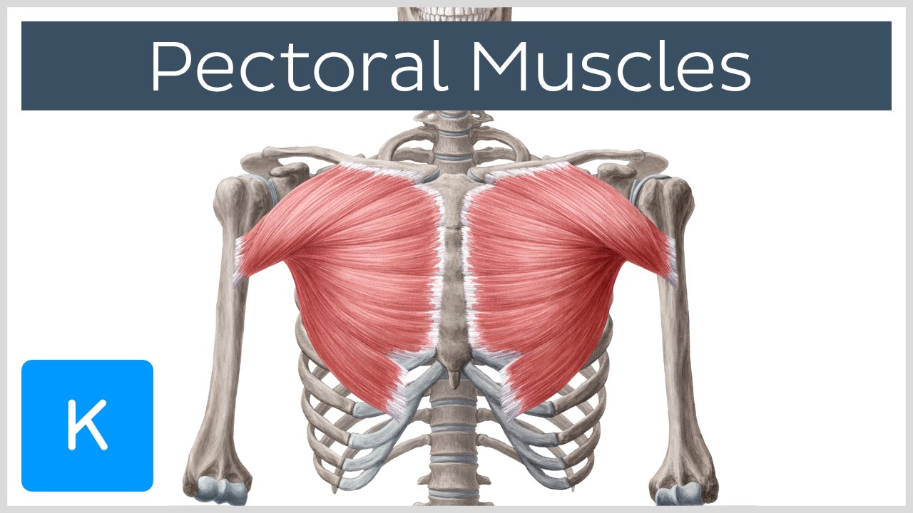
The chest anatomy includes the pectoralis major, pectoralis minor & serratus anterior. The epidermis is the outermost layer that provides a protective, waterproof seal over the body. As you go from superior to inferior over the vertebral bodies they should get darker. 12 photos of the anatomy of the chest area. Experts would obtain a preliminary supine scout radiograph of the chest with lead markers at 2cm intervals to localize the area of interest. Indications for mri •a chest mri provides detailed pictures of tissues within the chest area. The bottom of the manubrium forms a joint with the uppermost portion of the body of the sternum known as the manubriosternal joint. Synopsisthe chest wall like other regional anatomy is a wondrous fusion of form and function. Female reproductive organs front view. Find the perfect chest anatomy stock photo. Normal anatomy a bump on anterior sternum located beside 2 rib in center of chest.i cant find the name anywhere? Identify the basic anatomy seen on a chest radiograph. Chest pain is in mid section of chest and to the left.
The major muscle in the chest is the pectoralis major. An opacity (a normally dark area appears light on the image) a density (a. Gurmukh singh answered 49 years experience pathology sternal angle: The pectoralis major, pectoralis minor, serratus anterior and subclavius. Anatomy of chest area :
Pectoral Muscles Area Innervation Function Human Anatomy Kenhub Youtube from i.ytimg.com As you go from superior to inferior over the vertebral bodies they should get darker. Human anatomy for muscle, reproductive, and skeleton. Most chest bone fractures are due to trauma from a car accident. Huge collection, amazing choice, 100+ million high quality, affordable rf and rm images. Identify the basic anatomy seen on a chest radiograph. This is the most common section noted in sternum fractures. The epidermis is the outermost layer that provides a protective, waterproof seal over the body. The best upper chest workout will.
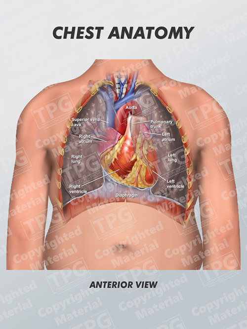
In particular, the right side of the chest is home to several structures including the right side of the heart, the three lobes of the right lung, the ascending aorta, the pulmonary blood vessels,.
Here, we break down the anatomy of your chest muscles. The pectoral region is located on the anterior chest wall. The pectoralis major and the pectoralis minor, known collectively as your pecs. Chest pain is in mid section of chest and to the left. Chest a man's chest — like the rest of his body — is covered with skin that has two layers. Fractures in the manubrial area are the second most common. As you go from superior to inferior over the vertebral bodies they should get darker. General anatomy neuroanatomy head and neck anatomy thoracic anatomy abdominal and pelvic anatomy spinal anat. Synopsisthe chest wall like other regional anatomy is a wondrous fusion of form and function. The abdomen (commonly called the belly) is the body space between the thorax (chest) and pelvis. Chester chest with peripheral port access arm. The great veins, the superior and inferior venae cavae, and the great arteries, the aorta and pulmonary trunk, are attached to the superior surface of the heart, called the base. These areas are also known as the hidden areas.
Identify the basic anatomy seen on a chest radiograph anatomy of chest. Anatomy of chest area :


0 Komentar